
🤯 INCRÍVEL: 72 Remarkable Microscopic Pictures Showing Things Up-Close 😲
Grab your lab coat, or at least pretend you have one, and prepare to zoom way, way in because we’re about to explore a world that’s been hiding right under your nose this whole time. Ever wondered what a grain of pollen looks like up close? Or why everyday objects start looking like alien landscapes the second you stick them under a microscope?
Luckily for us, curious minds have been busy capturing this hidden universe and sharing the results online. Turns out the tiniest things can be the most mind-blowing, so buckle up, because these insanely cool microscope pictures are about to make you question everything you thought you knew about the small stuff.
Click here & follow us for more lists, facts, and stories.
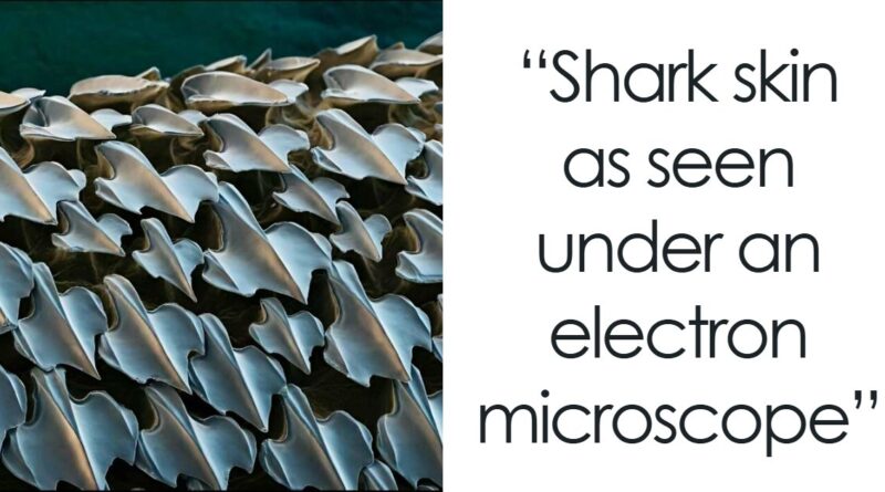
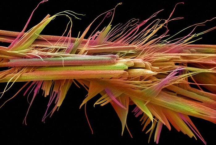
#1 Shark Skin As Seen Under An Electron Microscope
© Photo: Resident-Stage-3759
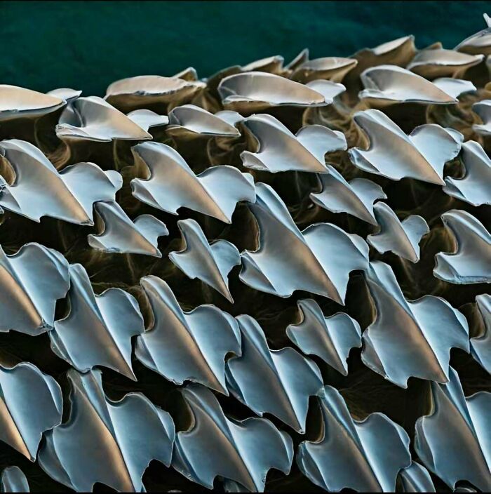
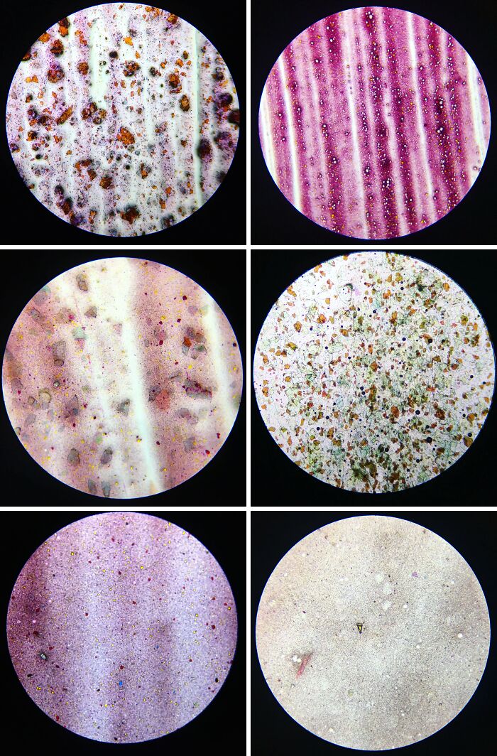
#2 Sand Under A Microscope, Magnified Up To 300x
© Photo: SnooLemons7574
Microscopes open a world invisible to the bare eye, bending light, or electrons, through lenses to enlarge tiny structures. Magnification makes objects bigger, while resolution determines how clearly fine details can be seen. Motic Microscopes explains that these principles are the foundation for everything we’re about to explore.
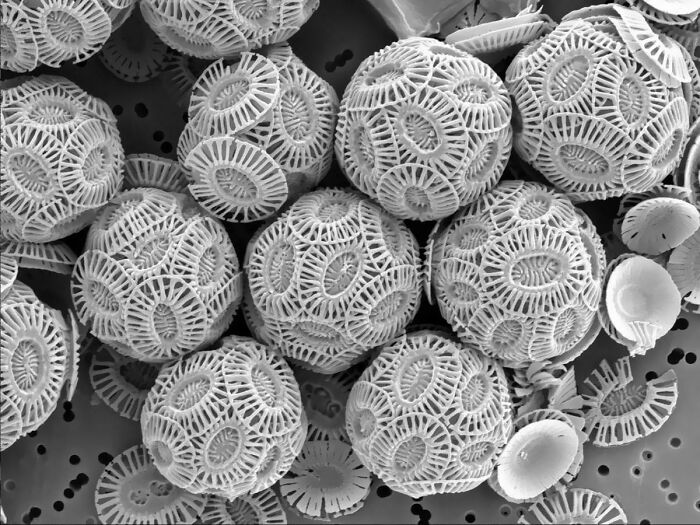
#3 Light Microscopy Image From A Skeleton Of A Diatom Algae 32 To 40 Million Years Old
“Mesmerizing light microscopy image from a skeleton of a diatom algae 32 to 40 million years old. Diatoms are photosynthesizing algae at the base of the marine food chain, found in almost every aquatic environment. They are single celled organisms that produce an external wall composed of silica. When they die, their silica shells accumulate on the floor of the body of water in which they live. Thick layers of these diatom shells have been fossilized into sedimentary rock called diatomite, or Diatomaceous earth!” – OCR
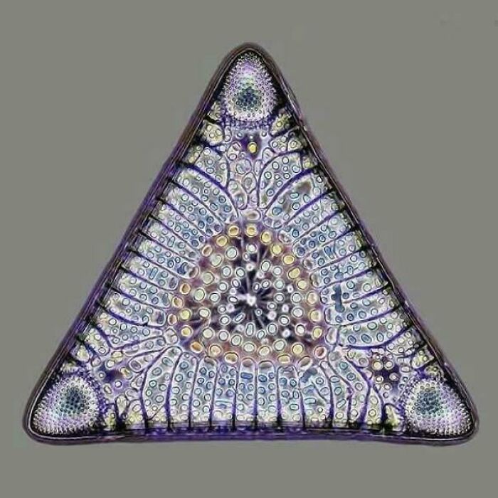
© Photo: Academic_Job_4665
#4 Chalk Particles Under A Microscope
© Photo: Knight_Fisher61
Different microscopes reveal different aspects of the microscopic world. Light microscopes allow scientists to view different specimens, often thinly sliced or mounted in liquid, while electron microscopes require non-living, dehydrated and sometimes metal-coated samples, producing extreme detail at the cost of life. Knowing the difference helps explain why some images look so alien compared to others.
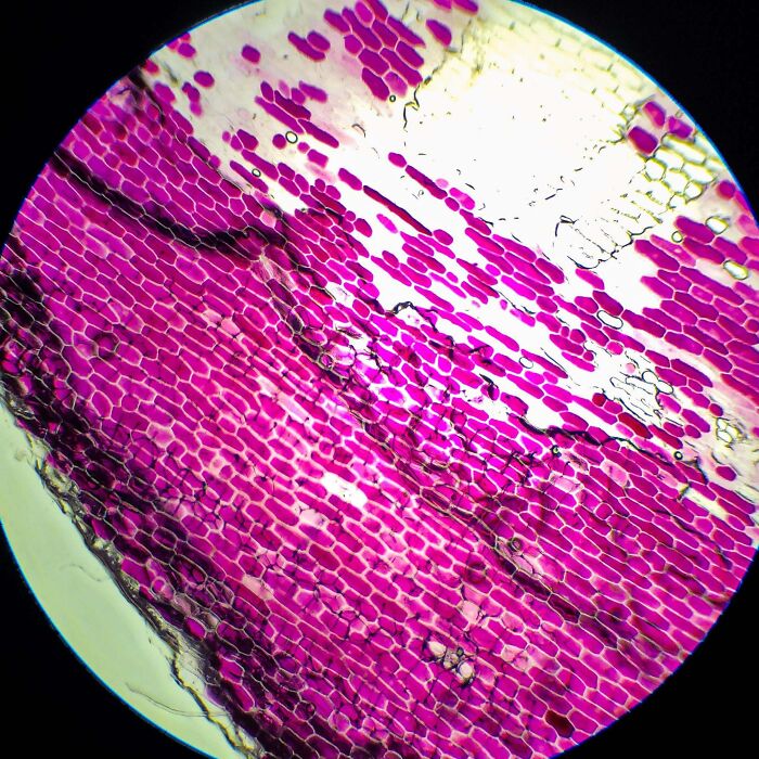
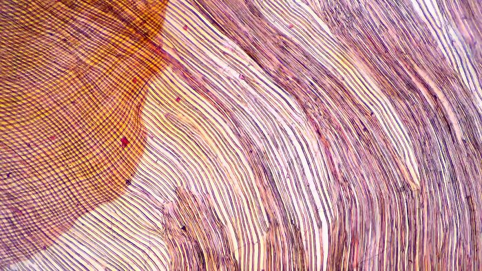
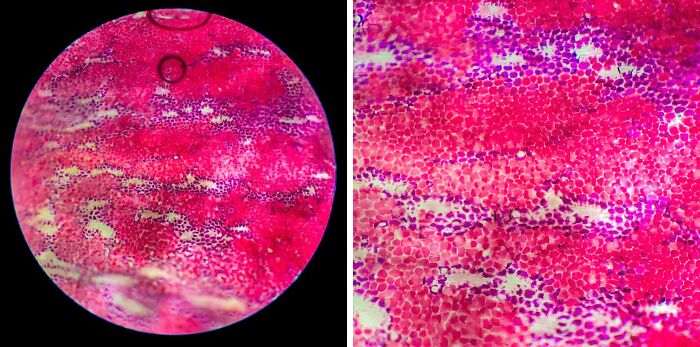
#5 Wood Under A Microscope Is Breathtaking
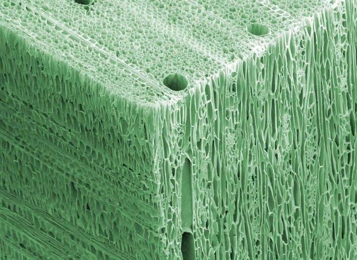
© Photo: reddit.com
#6 A Cat’s Tongue Under A Microscope Looks Like It’s Made Of Other Smaller Tongues
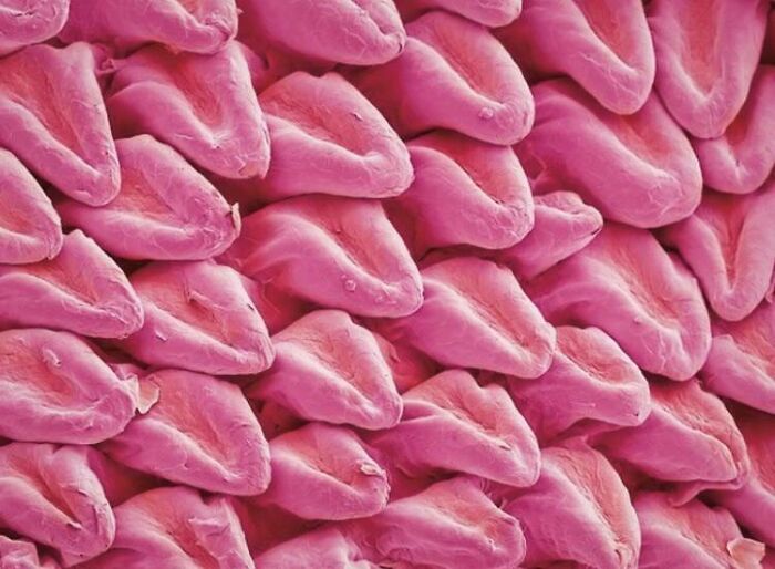
© Photo: Katica123
Milne Library adds that many microscope images look vividly colorful not because they naturally are, but due to staining techniques or digitally added false colors. Electron microscope images are originally black-and-white, so any color is applied afterward for contrast or visual appeal, creating almost artistic results.
#7 Cancer Cells Under An Electron Microscope
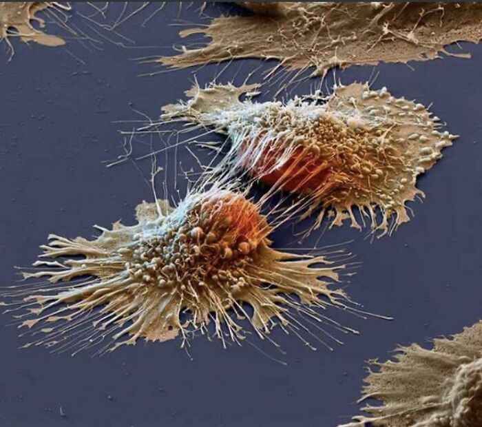
© Photo: User
#8 A Microscopic Look At The Tiny Creatures Living In Less Than A Millilitre Of Seawater
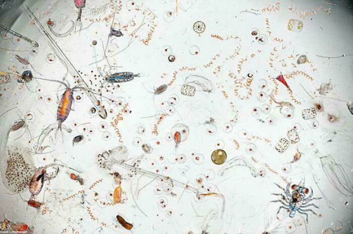
© Photo: r_person
Further explaining this, microbiologists and histologists routinely use dyes to make transparent cells visible. Simple stains color entire cells, while differential stains, like Gram or acid-fast staining, highlight specific structures or species. For example, Gram-positive bacteria appear purple, while Gram-negative bacteria turn pink, helping scientists differentiate types of organisms under the lens.
#9 Pollen On A Horse Fly’s “Eyelashes”
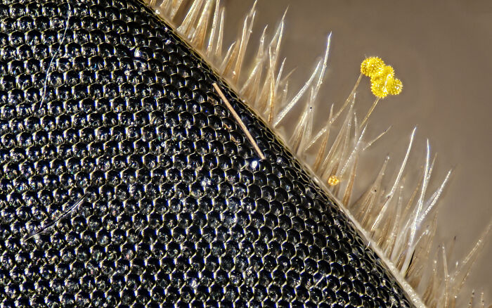
© Photo: DoYouTasteMetal
#10 Plasmolized Red Onion Cells Under Microscope
© Photo: doctorchibanga
Earthly Mission explains that microscopes open the door to a hidden world that looks completely different from our everyday experience. Objects that seem smooth, simple, or even boring to the naked eye suddenly reveal complex textures, intricate patterns, and entire ecosystems of microscopic life. A plain surface can turn into a rugged landscape, and something lifeless can appear full of structure and activity.
#11 Grass Under A Microscope
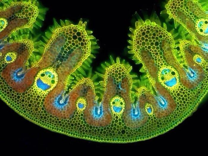
© Photo: HBeel
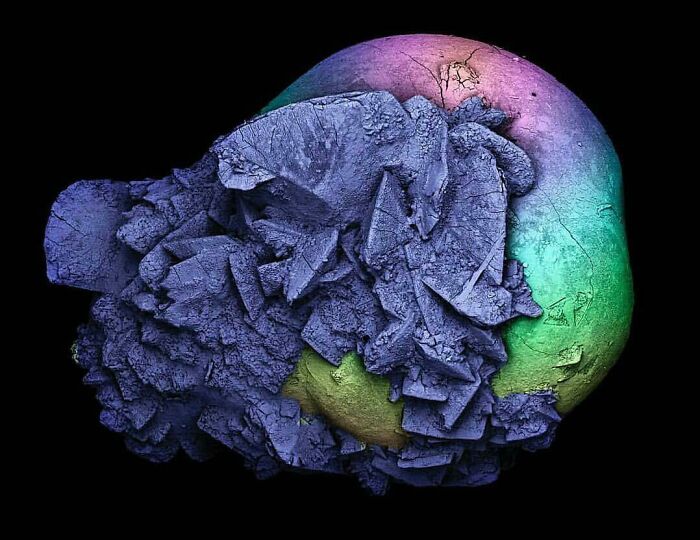
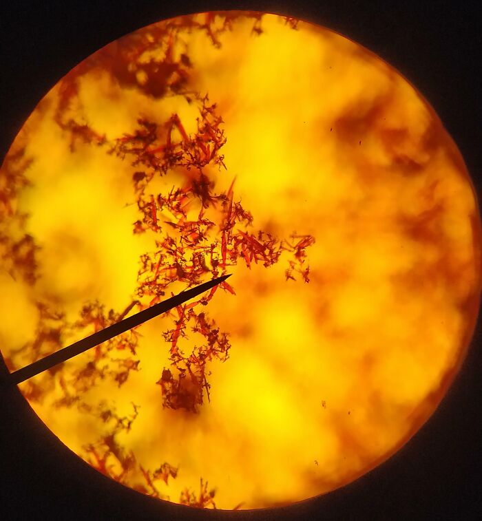
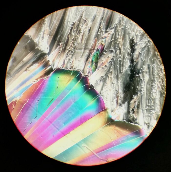
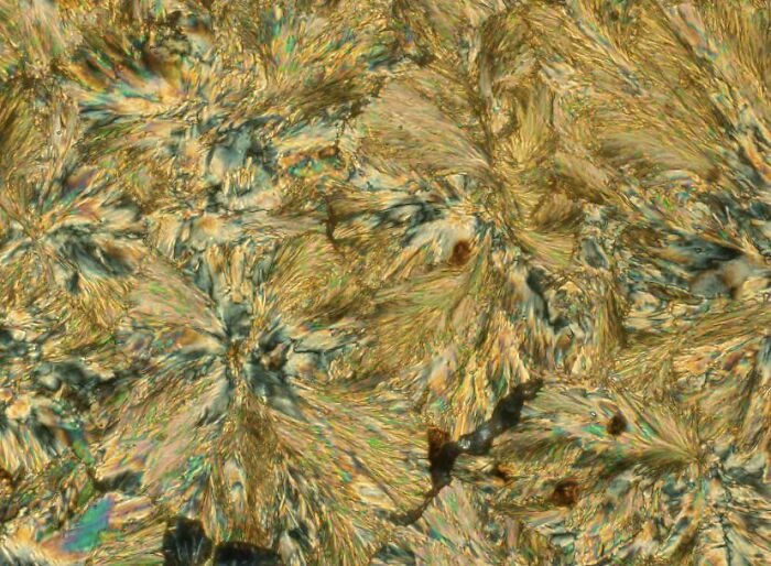
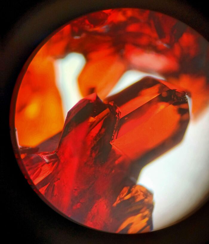
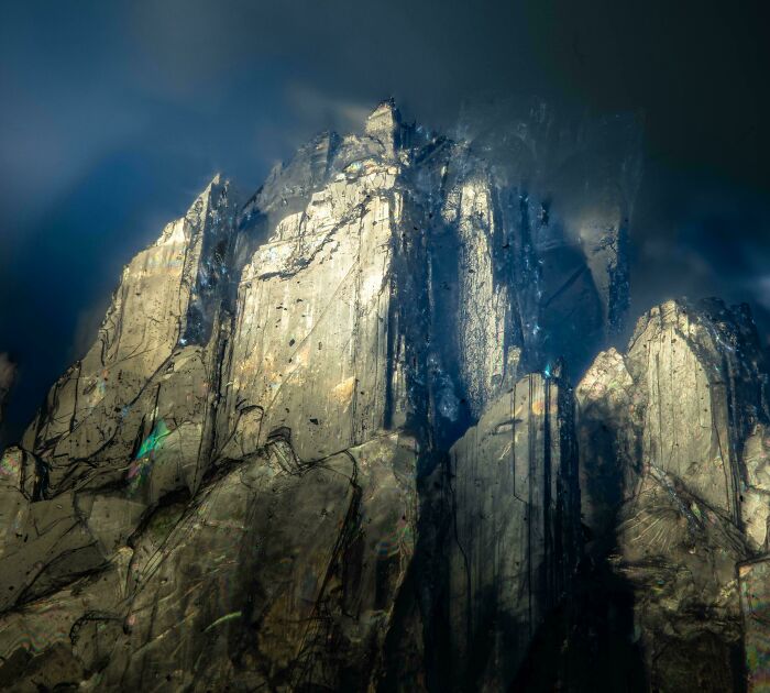
#12 This Microscopic Mountain Range Is A Combination Of Crystallized Lidocaine And Gentisic Acid In Ethanol
40x total magnification, shot with an iPhone camera.
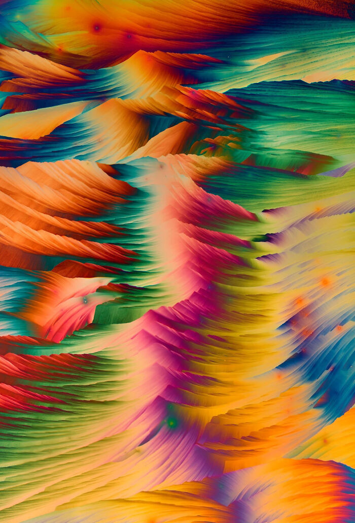
© Photo: Martinkaae
Under magnification, familiar items take on entirely new forms. Bacteria appear as rods, spheres, or spirals on surfaces we consider clean. Insects show intricate compound eyes and jointed limbs, while salt crystals reveal sharp-edged cubes. Hair shows layered textures, and fabrics expose tangled fibers often carrying dust or microbes.
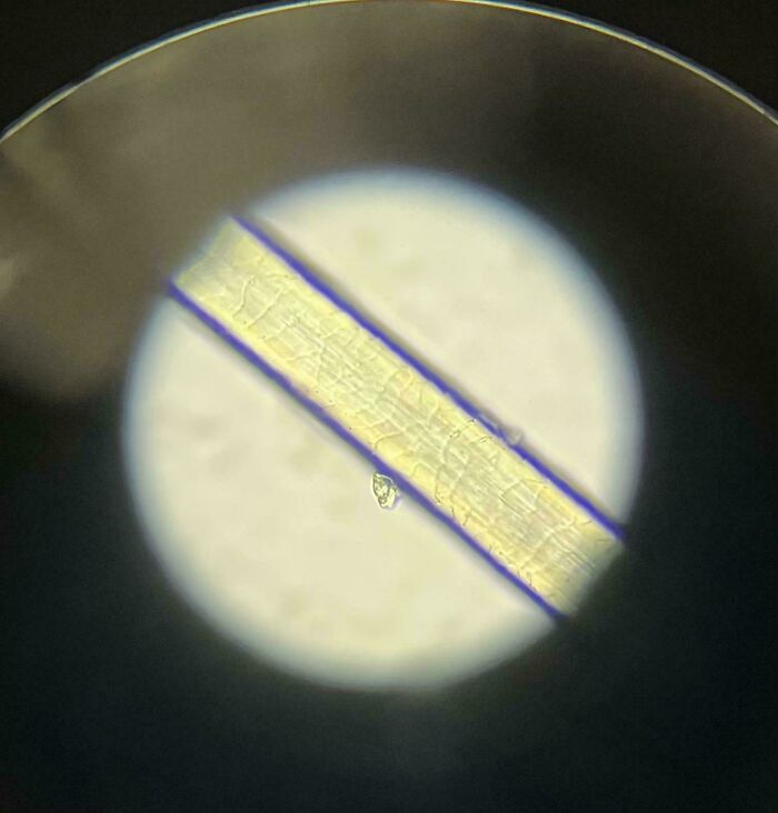
#13 New Ballpoint Pen Under The Microscope
© Photo: dubhead_dena
#14 Sitting In Bed, Felt A Bite, Saw This Little Guy Crawling On Me. Threw Him Under The Microscope
© Photo: Vif-Argent
DSS Image explains that microscopy is more than just visually striking pictures, it is essential for science, medicine, and materials research. Observations under the microscope inform medical diagnoses, drive scientific discoveries, and guide technological and quality-control advances.
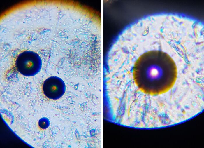
#15 Picture Of Saccharin In My Polarising Microscope
© Photo: 8thunder8
#16 Kidney Stones Under A Microscope. Size 2 Mm
© Photo: dubhead_dena
In practice, hospitals and labs use microscopes to detect infections, cancer, and blood disorders by examining cells, tissues, and microbes. They help identify pathogens, monitor tumor grading, and support treatment decisions. Similarly, materials scientists use microscopes, especially SEM, to study tiny defects, cracks, and grain structures in metals and composites, improving their performance and safety.
#17 Took Some Pictures Of Chloroplasts Under The Microscope
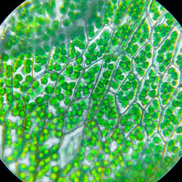
© Photo: doctorchibanga
#18 The Colloidal Iron Tissue Stain Utilizes Prussian Blue Chemical Reaction To Stain Acidic Mucin Secretions With Prussian Blue Color For Viewing Under A Microscope
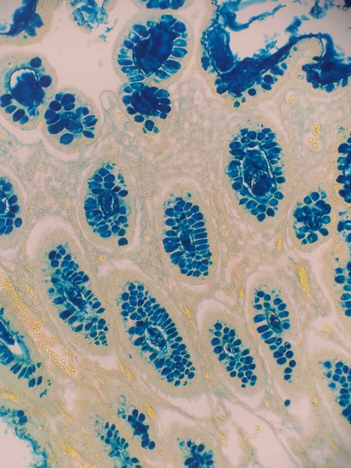
© Photo: reddit.com
Equinecare Probiotic shares that microscopy also reveals some astonishing details: standard light microscopes can magnify up to 1,500×, turning dust into jagged landscapes, pollen into glowing miniature objects, and mold into forests of branching filaments reminiscent of alien landscapes.
#19 Robber Fly Wing
I don’t shoot photos using a microscope, but a mirrorless camera with a custom built tube lens and various microscope objectives. My magnification range for images, is 10x to 40x. Sony A7R3 camera body. Tube lens using a reverse mounted Raynox DCR-150. 10x/20x/40x Mitutoyo objectives. 300 watt strobes (x2). 3 axis camera and subject positioning with sub micron resolution. 1200 pound scientific/precision granite block table on vibration isolating feet.
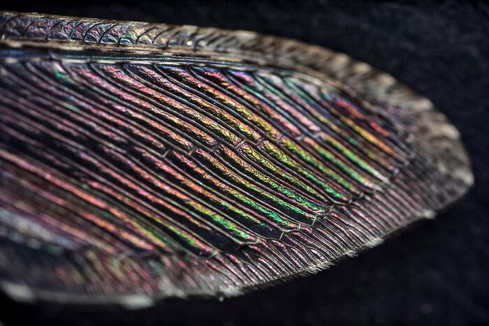
© Photo: BPLEquipment
#20 Pollen From A Bees Leg Under A Microscope
© Photo: kooperkape
Looking closer at biology, human hair in cross-section shows rings and cracks like tree rings, while split ends appear frayed. Cardiac and muscle tissues form mosaic-like patterns, and red blood cells observed with electron microscopes appear as smooth, dimpled discs rather than flat circles. These details highlight the unexpected complexity in the familiar.
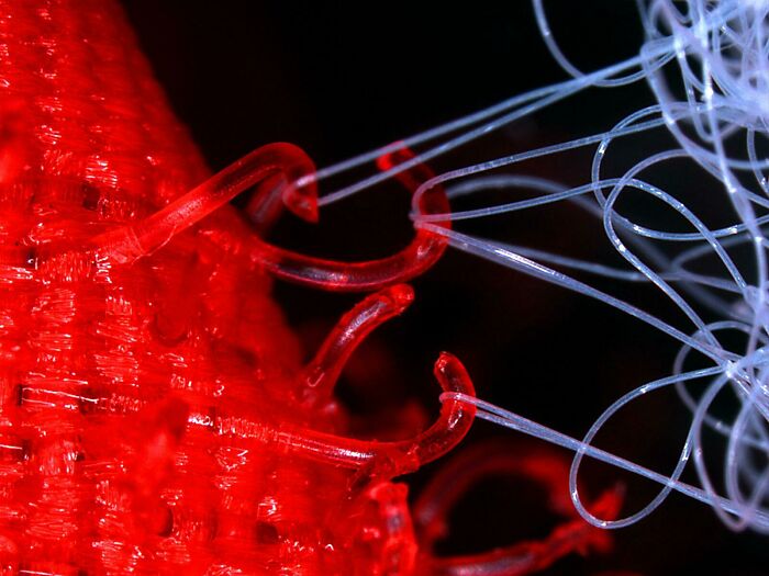
#21 Polyp Lifestage Of A Jellyfish (Obelia Sp., Hydrozoa, Cnidaria) I Investigated Under The Microscope. Also Called “Sea Fur”
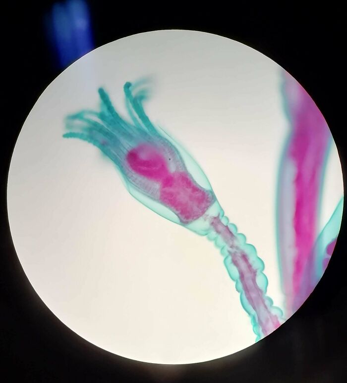
© Photo: nureinpanda
#22 When You Look Into The Microscope And Something Looks Right Back At You
© Photo: User
Finally, some microscopic animals, like loriciferans, are smaller than many single-celled organisms but still possess a brain, gut, and reproductive organs. At the atomic scale, advanced microscopy can even visualize individual atoms or layers just two atoms thick, offering a glimpse into the building blocks of matter itself.
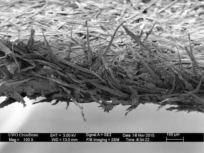
#23 A Used Toothbrush Bristle Under An Electron Microscope
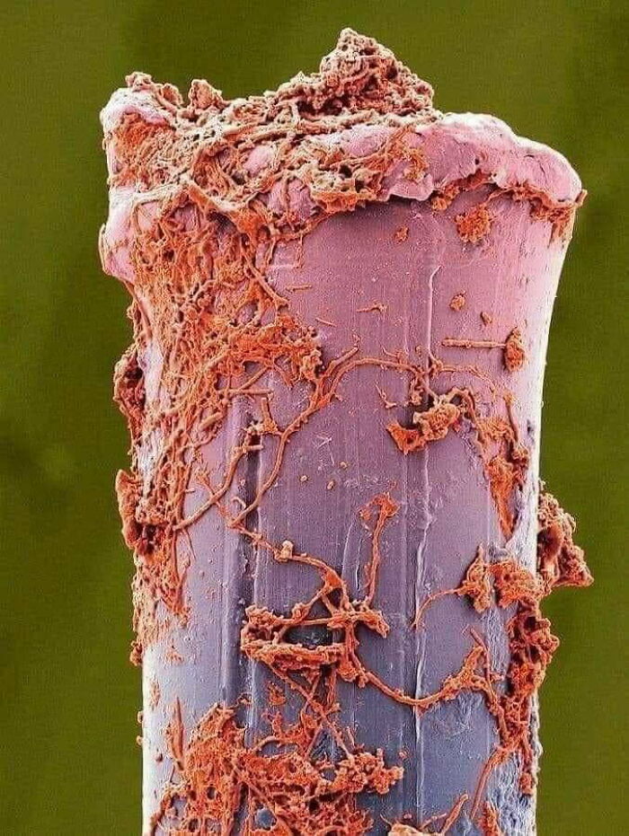
© Photo: trisaders
#24 Caffeine Crystals Under A Microscope
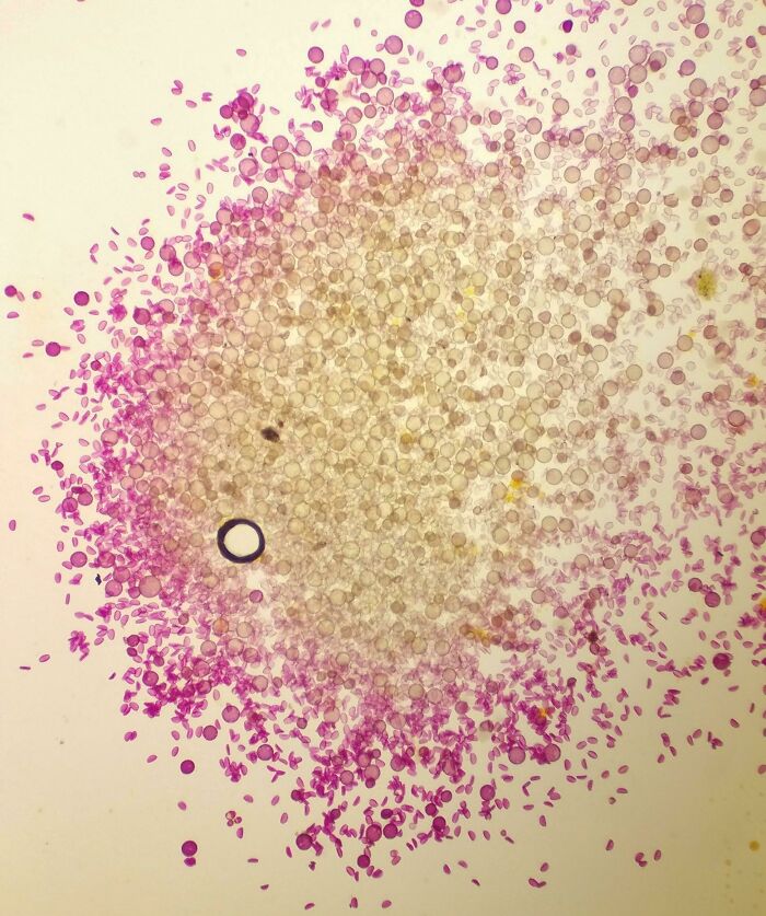
© Photo: User
At the heart of all these mesmerizing images, microscopy isn’t just about making tiny things look bigger, it’s about changing the way we see the world entirely. What seems ordinary at first glance can turn into something extraordinary with just a little magnification, reminding us that there’s far more detail hiding beneath the surface than we ever notice in our day-to-day lives.
Of course, not all microscopic discoveries are purely aesthetic, many play a role in science, medicine, and our understanding of life itself. However for the rest of us, they offer something just as valuable such as a fresh perspective. So the next time you glance at something as simple as a strand of hair or a grain of salt, remember you might just be looking at something far more fascinating than it seems.
#25 Sem Images Of Zinc Oxide Nanowires I Did During My Master Thesis
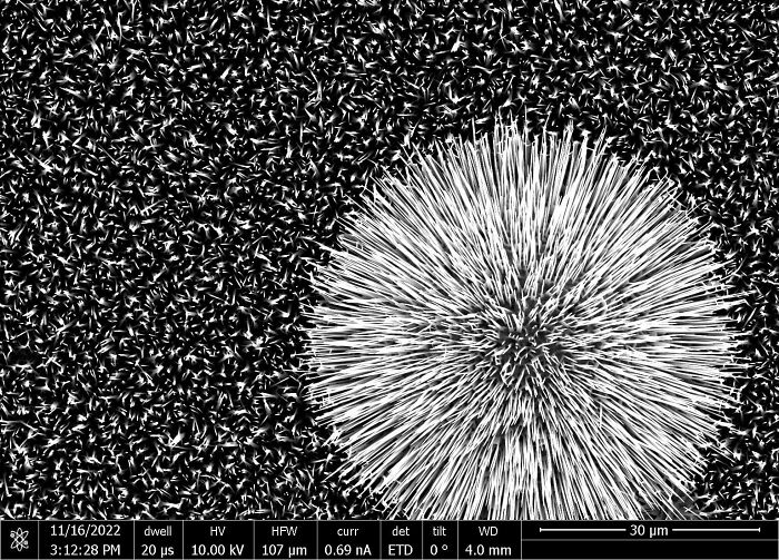
© Photo: TemporarySun314
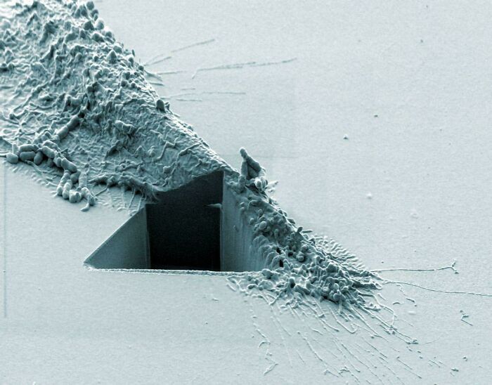
#26 View Of Torn Paper Under The Microscope
© Photo: nanofabrication
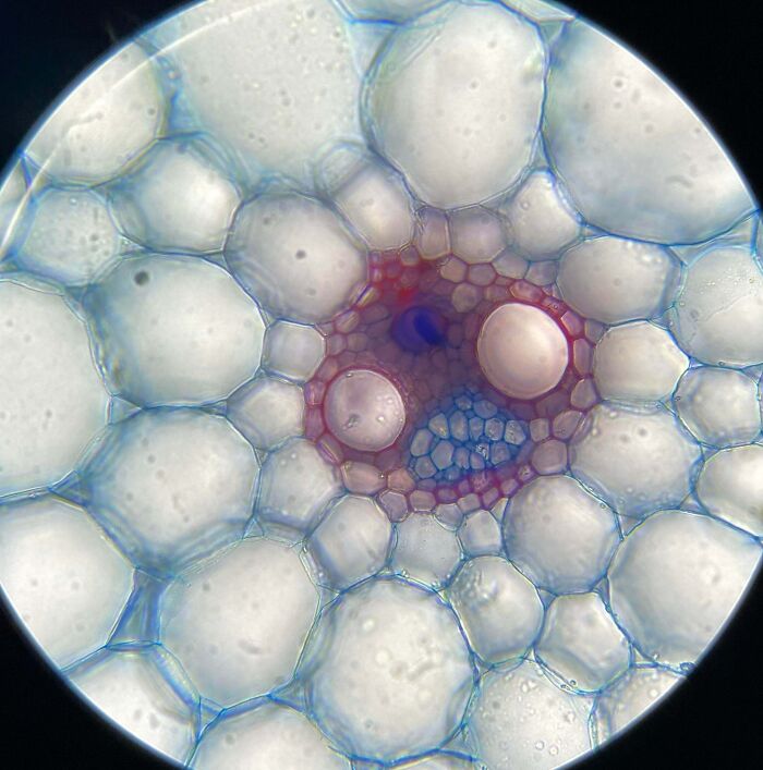
#27 Bud Cut From A Cherry Tree
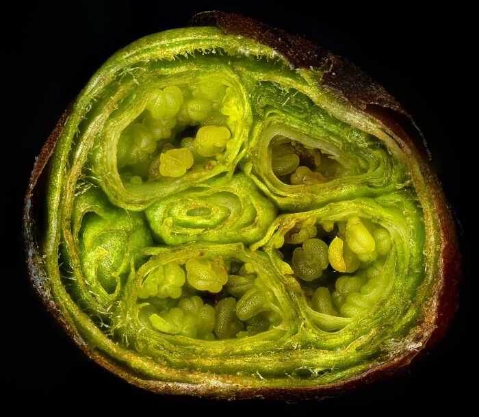
© Photo: Alexander_Klepnev
#28 Microscopic Dust Particles Under An Electron Microscope Resembles Cosmic Collisions
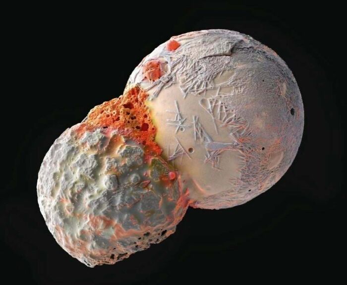
© Photo: Scientiaetnatura065
#29 I Found A Snake Shed And Put It Under My Microscope
© Photo: Express_Buffalo7118
#30 Microscopic Picture Of A Guitar String
© Photo: Rattlesnake_Mullet
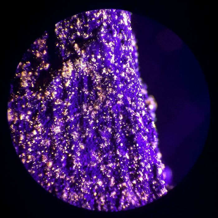
#31 Silver Dichromate Crystals Under The Microscope
© Photo: CLoncha
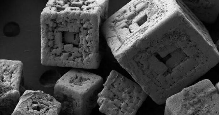
#32 I Took A Picture Of Sugar Under A Microscope
© Photo: Sofabezug
#33 Winged Seed Under Microscope
© Photo: JayDu1981
#34 This Is What Velcro Looks Like Under A Microscope
© Photo: Niyi_M
#35 Table Salt Under A Microscope Looks Surreal
© Photo: reddit.com
#36 I Compared Some Shades Of Lipstick Under The Microscope
MUA Shimmer Lipstick in Rose Gold, Wet N Wild Liquid Catsuit in Berry Recognize, Tarte Lipsurgence Lipstick in Adored, ULTA Sweet and Shimmer Lip Gloss, Tarte Lipsurgence Lipstick in “Decadence”, Tarte Tarteist Lip Paint.
© Photo: phnx0221
#37 I Took Some Pictures Of A Rose Under The Microscope
© Photo: doctorchibanga
#38 A Picture I Took Of A Liquid Crystal Under The Microscope. The Same Type Of Molecules That Are On The Displays Of Computers
© Photo: freelafrancener
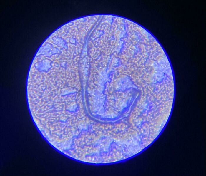
#39 Freshwater Flatworms Under DIC Microscope
© Photo: VegetableDiscount517
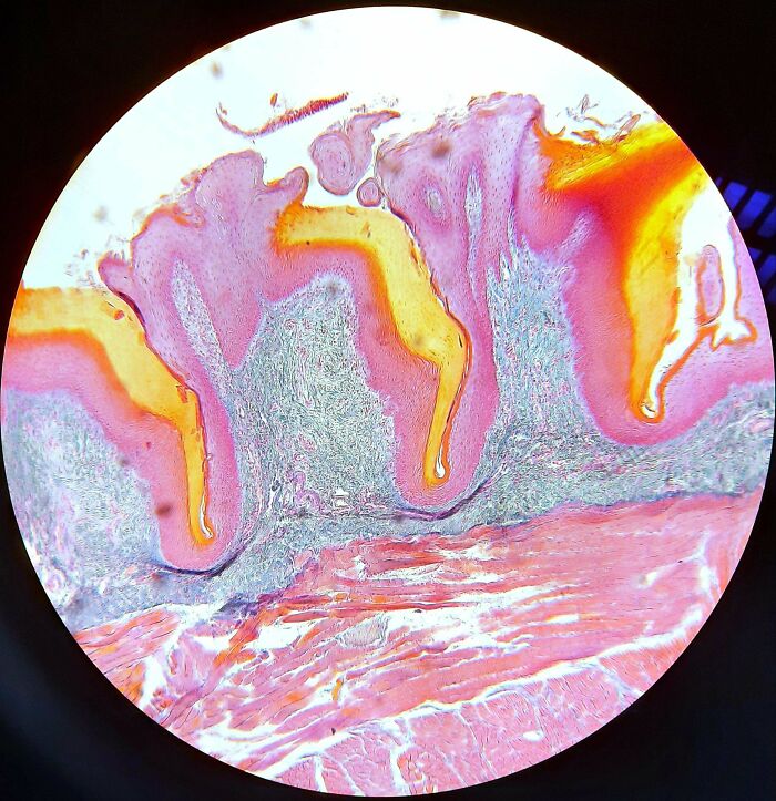
#40 We Had To Work With 25 Year Old Samples, And If I Am Correct This Is A Tongue. I Find It Insane That Prepared Samples This Old Still Look This Goof Under A Microscope!
© Photo: justslightlyodd
#41 A False-Color, High-Res Microscopic View Of Prions
Prions are rogue, misfolded proteins that arise in the brain and cause neurodegenerative diseases with a 100% fatality rate. They can even be transmitted between people.
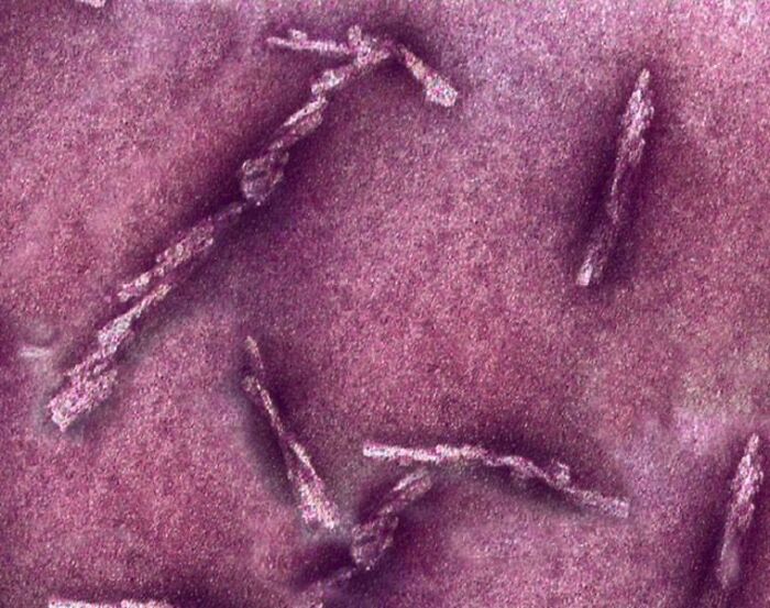
© Photo: JurassicPark9265
#42 Took A Photo Of A Red Pepper Flake Through A Microscope And It Looked Like A Planet
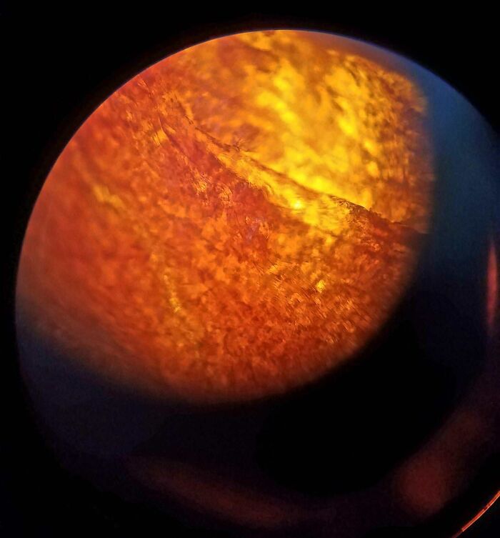
© Photo: imgur.com
#43 Got Bored During Class And Put Small Pieces Of Used Eraser Under A Microscope After I Finished My Lab
© Photo: User
#44 White Hair Is Actually Transparent (Taken With A Microscope, White Hair In Front Of A Regular Hair)
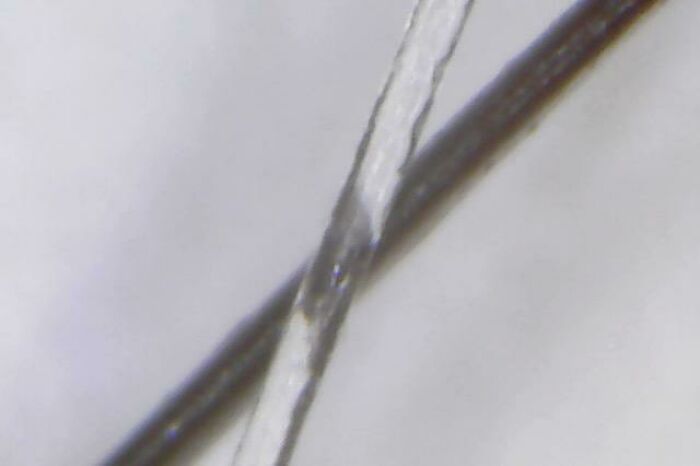
© Photo: lau1159
#45 A Cancer Cell Slashed Open By An Ion Beam
© Photo: reddit.com
#46 Some Plants And Red Algae Under The Microscope
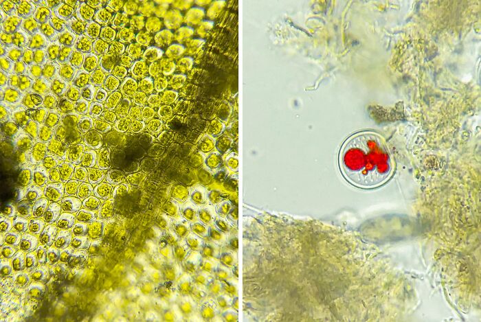
© Photo: doctorchibanga
#47 Extremely Fluorescent Crystals Of Derivatized Dibenzalacetone Under Microscope
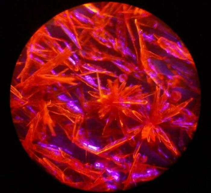
© Photo: m_ichor
#48 Potassium Ferric Cyanide Under The Microscope
© Photo: Niklas_Science
#49 The Semicrystalline Nature Of Vaseline And A Protic Ionic Liquid
I smeared vaseline and a surfactant (triethanolamine oleate) on glass slides. Then I performed cross-polarized optical microscopy to obtain these images. GPT enhanced the vibrancy of the blues. The blue colors result from birefringent interference. Notice the fibrous needle morphology in the vaseline compared to the lyotropic lamellae of the triethanolamine oleate.
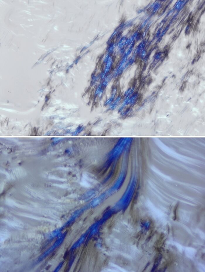
© Photo: naftacher
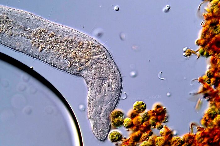
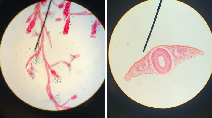
#50 Found This Worm On A Slice Of Onion In Biology Class, Can Only Be Seen Under A Microscope
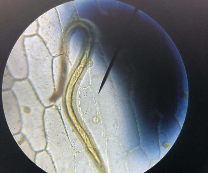
© Photo: emmy_the_egg
#51 Interesting Arrangements Of E. Coli Under Microscope
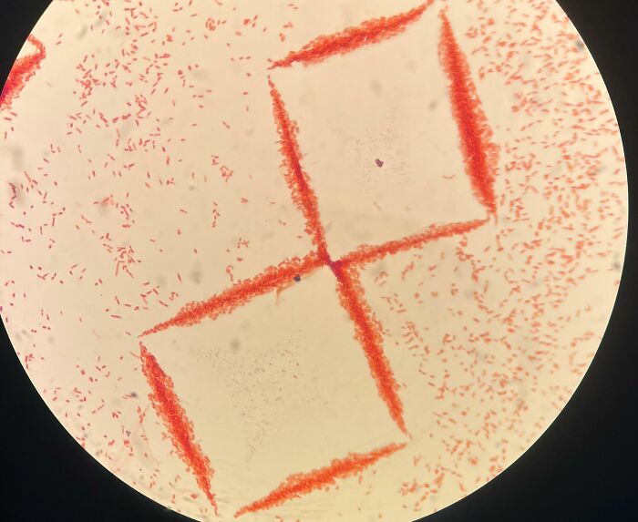
© Photo: JokellOwO
#52 You Can See Hair Toner Under A Microscope
© Photo: blanket_shark
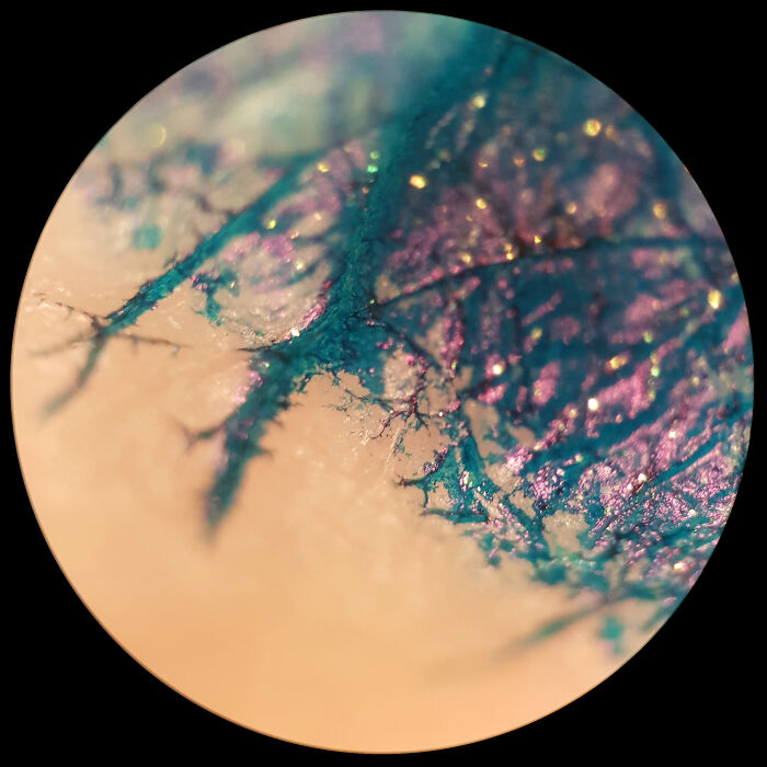
#53 Fluorescent Spray Paint On Stick, 40x
© Photo: yooooooUCD
#54 Microscopic Cross-Section Of The Leg Of A Red Centipede
© Photo: DrawingwithJB
#55 Ink On Skin Under The Microscope
© Photo: RightyHoThen
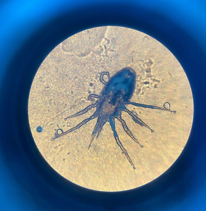
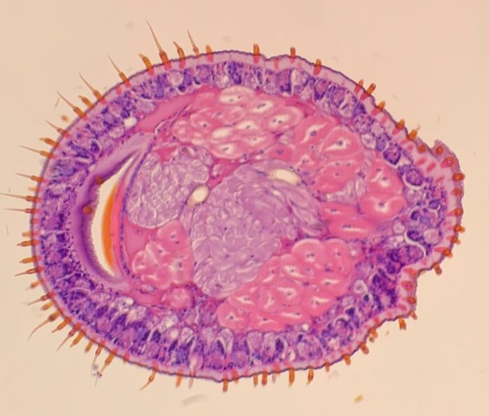
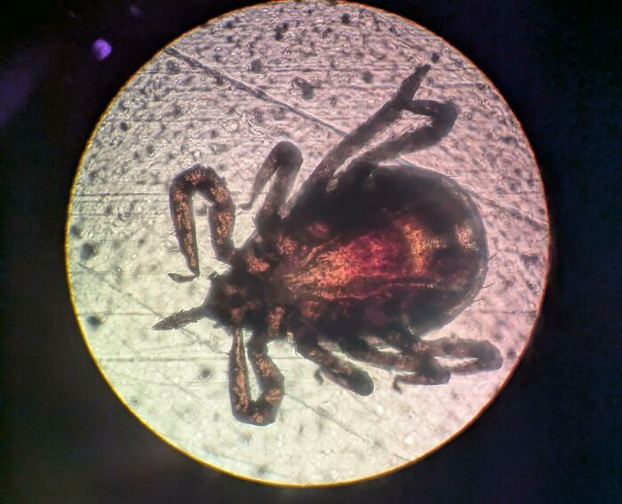
#56 A Paralysis Tick Under A Microscope
© Photo: Spnvettech
#57 Musculoskeletal Tissue Lab Practical-Some Pics From The Microscope
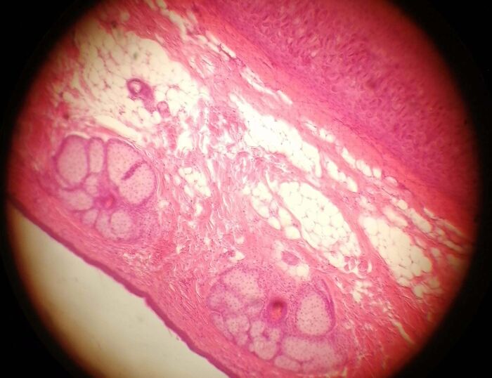
© Photo: 4Ever-A-Stick-insect
#58 Electron Microscope Images Of The Human Lip, A Shaved Hair And A Nail Bed
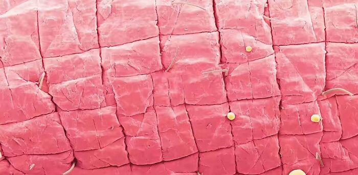
© Photo: Certain-Dark-3955
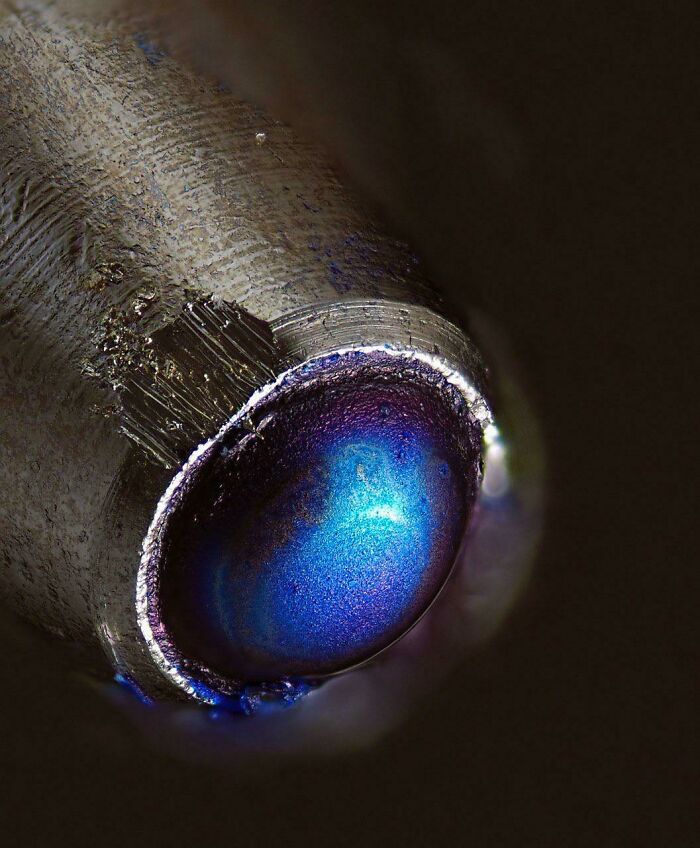
#59 Vanadium Hexa-Aqua Under A Microscope
This type of vanadium had a beautiful rainbow-like color on it. It looks beautiful both under the microscope and in hand.
© Photo: ChemBroDude
#60 Was Playing Around With My Son’s New Microscope. Decided To Look At A Drop Of My Blood
© Photo: Ace-Of-Mace
#61 Kyanite Under A Microscope
© Photo: pen_n_paper
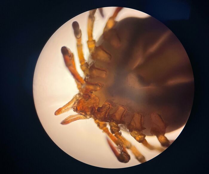
#62 Removed A Tick
© Photo: Philosophical_Sayer
#63 I’m Taking Zoology And I Thought I Would Share Some Of The Stuff I’ve Looked At Under The Microscope
© Photo: livkons
#64 Got A New Microscope For Soldering And My Dad Still Had Some Fresh Greens From His Vegetable Garden
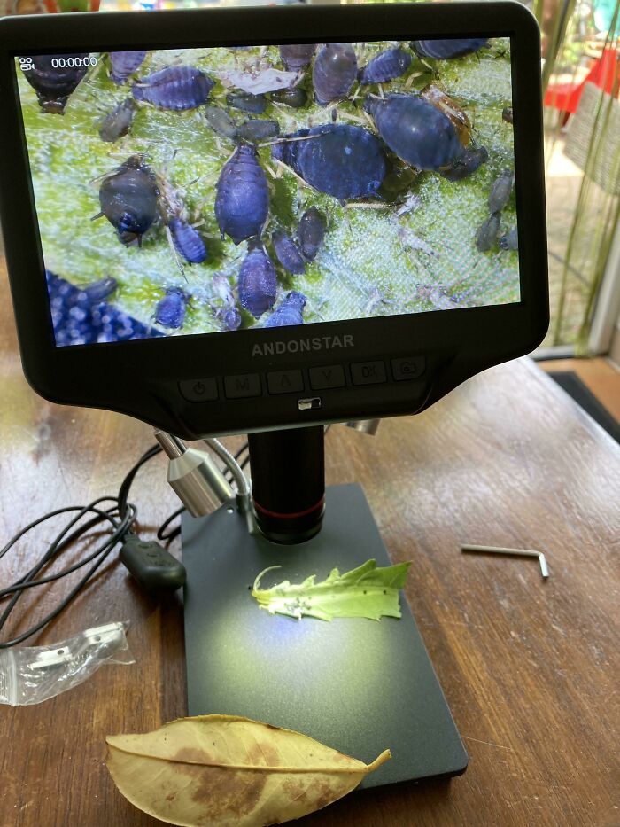
© Photo: captainkanpai
#65 Perfect For The Spooky Month, Jack-O-Lantern Neutrophil
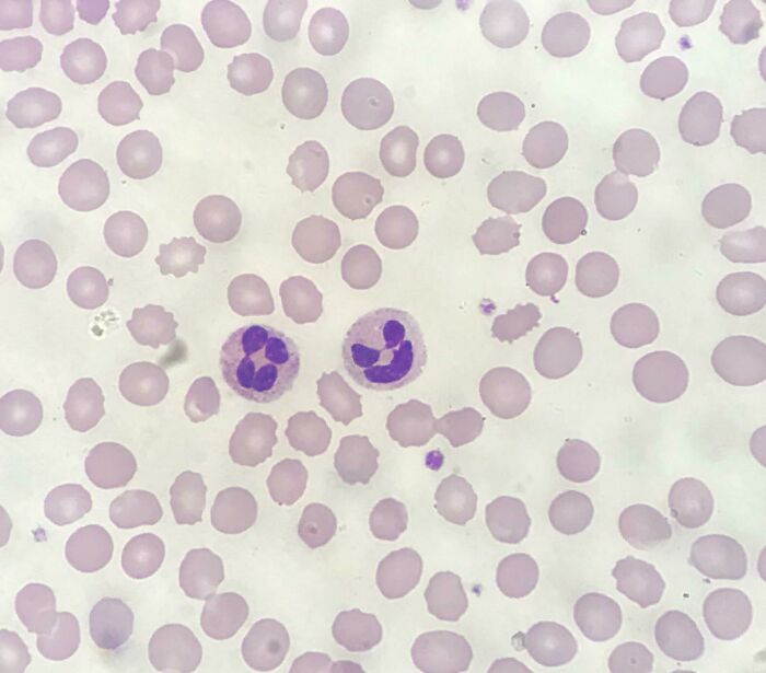
© Photo: User
#66 Two Peices Of Sapele Wood Glued Together And Under A 30x Microscope
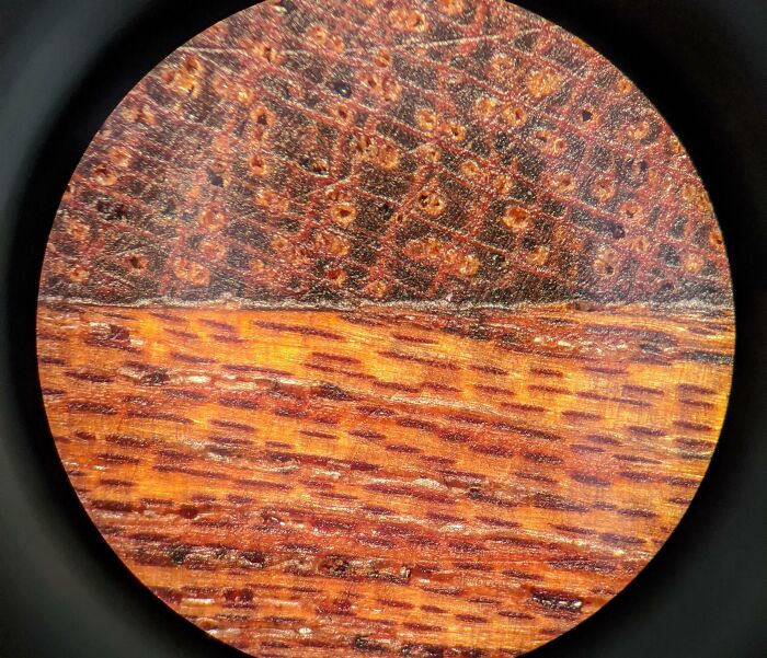
© Photo: DesTaches
#67 My Saliva Through My Sons Microscope
© Photo: thedemocracyof
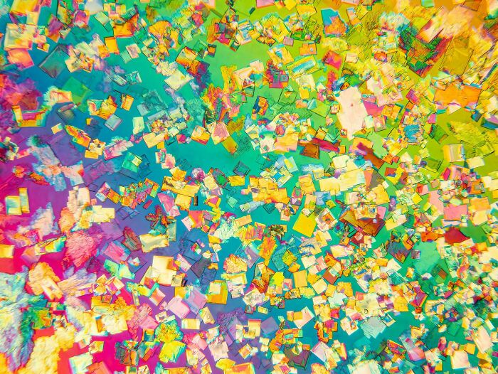
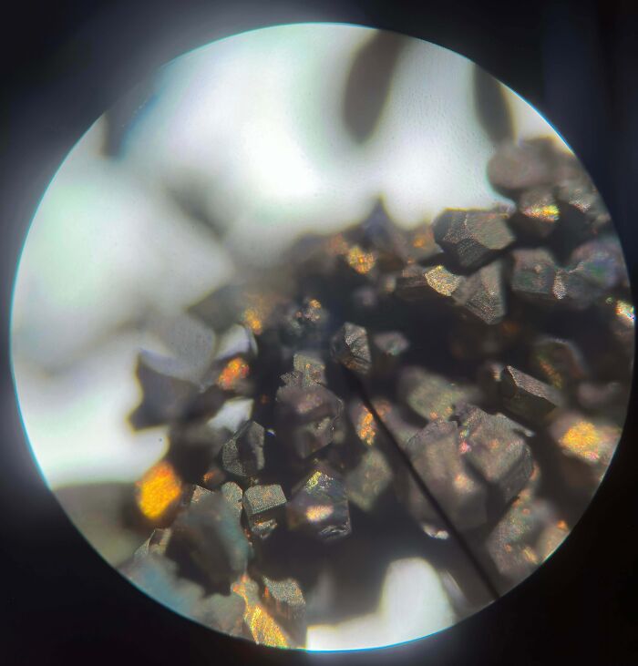
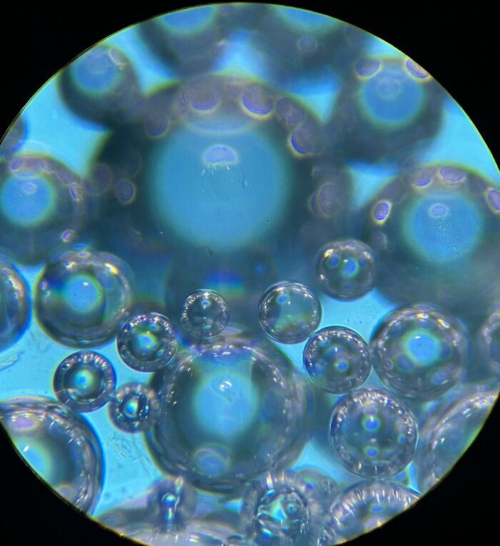
#68 Check Out The Perfect Geometry Of These Hydrate Resins Under Microscope
© Photo: Kijjy
#69 Here Is A Close Up Of A Misquote Larva Under A Microscope. Looks Vicious
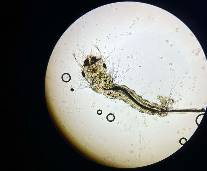
© Photo: bennolen
#70 Took A Picture Of Cat Ear Mites Under A Microscope

© Photo: okaythen1guess
#71 Some Caffeine I Extracted The Other Day Under Microscope
© Photo: TheSaucez
#72 Got A New Microscope And Had A Look At My Partners Saliva
© Photo: StuffedFerret
You might also like: 50 ‘Weird Facts’ About The World That Might Give You A Fresh Perspective
📢 Gostou da notícia? Compartilhe com os amigos!
Este artigo é uma tradução automática de uma fonte original. Para ler o conteúdo na íntegra: Clique aqui.